Multi-lead visualization
In this tutorial you will learn how to visualize several ECG channels within the PhysioZoo Peak detection module.
Introduction
PhysioZoo allows you to visualize several ECG channels in parallel. TThis can be use to facilitate the review of Holter recordings for example. This tutorial will show how to display several channels in parallel.
Displaying several leads
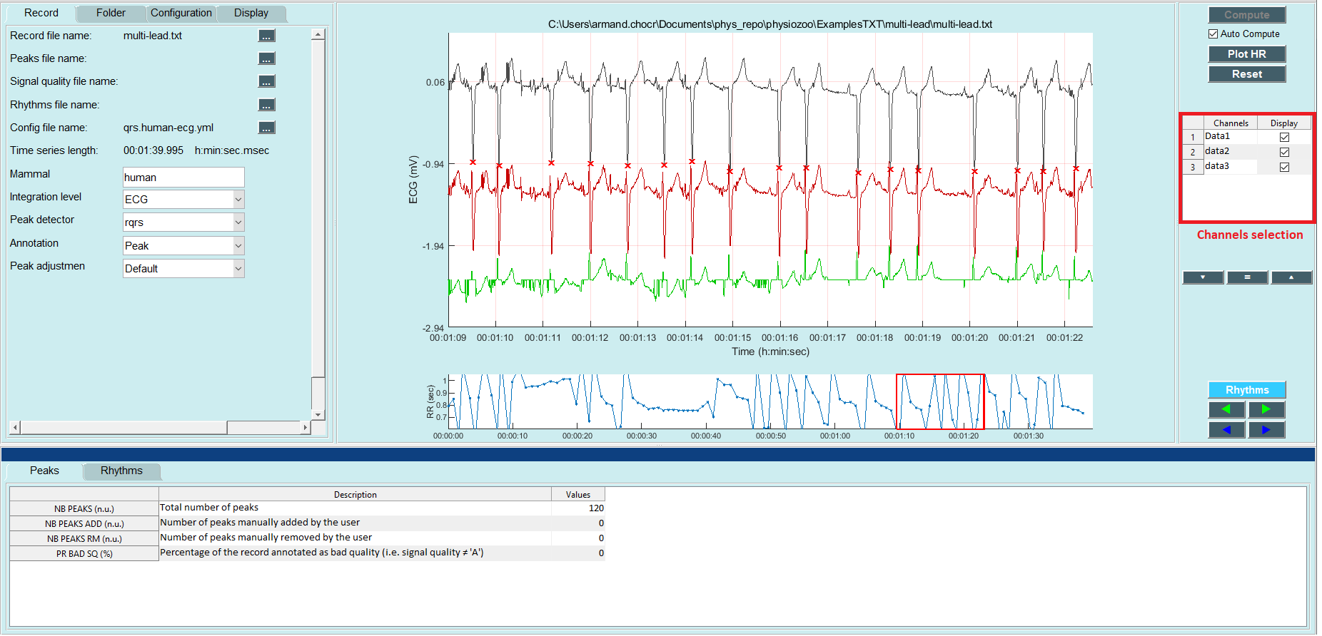
To display several channels of an ECG, follow these steps:
Select the multi-lead ECG example: File-> Open data file-> physiozoo\ExamplesTXT\multi-lead\multi-lead.txt.
On the right panel, select the desired channels to be displayed. By default, only channel 1 is selected.
Frequently asked questions
What is the format of the files to be loaded to **PhysioZoo to display several channels ?**
If recordings are provided in .mat format then each column will represent a different lead.
An other option is to use a .txt file which will contain the following header: